Diagram Of Shoulder Muscles And Tendons | Following inferior dislocation of shoulder joint, the rounded contour of shoulder is lost and there is weakness of abduction of armbecause the axillary nerve is likely to be injured in the inferior. They indicate swelling (inflammation) of a particular area within the the shoulder joint is kept stable by a group of muscles called the rotator cuff as well as the biceps tendon. The shoulder muscles include skeletal muscles that are attached to the head of the humerus which performs various direct and indirect functions of the both heads join to form one large muscle the tendon of which inserts into the radial tuberosity. The joint is strengthened and stabilized by adjacent muscles and tendons, especially by the musculotendinous rotator cuff. The clavicle (collarbone), the scapula (shoulder blade), and the humerus (upper arm bone) as well as associated muscles, ligaments and tendons.
Hold tendons of long head of biceps brachia muscles in groove between the greater and lesser tubercle on humerus. The anterior capsule is thickened by the three glenohumeral ligaments while the tendons these are the supraspinatus, infraspinatus, teres minor and subscapularis muscles. Related posts of shoulder muscles and tendons diagram muscles of the shoulder. We'll discuss the function and anatomy. Shoulder joint muscles (glenohumeral joint) the shoulder joint has very large powerful muscles which provide the power for strong movements in addition to shoulder dislocations, other common injuries include rotator cuff tendon tears and broken bones including the humerus and collar bone.

We'll discuss the function and anatomy. Know the anatomy of the shoulder involving its skeletal system, cartilages, ligaments, muscles, tendons. Once the ligaments, tendons, and muscles around the shoulder become loose or torn, dislocations can occur repeatedly. Diagram of shoulder tendons shoulder joint anatomyskeletal systemcartilagesligamentsmuscles. Shoulder joint muscles (glenohumeral joint) the shoulder joint has very large powerful muscles which provide the power for strong movements in addition to shoulder dislocations, other common injuries include rotator cuff tendon tears and broken bones including the humerus and collar bone. Muscle anatomy of the neck. They indicate swelling (inflammation) of a particular area within the the shoulder joint is kept stable by a group of muscles called the rotator cuff as well as the biceps tendon. Tendons are extensions of muscles that attach muscles to bone. For that reason, and because of the dexterity of the shoulder joint itself, the musculature of the shoulder is complex, ranging from massive prime mover muscles to. Printable shoulder muscles diagrams to help you study the muscles structure in human's shoulder.we have five muscle diagrams of the shoulder. This usually occurs secondary to repetitive use of the shoulder. The shoulder muscles play a large role in how we perform tasks and activities in daily life. The anterior capsule is thickened by the three glenohumeral ligaments while the tendons these are the supraspinatus, infraspinatus, teres minor and subscapularis muscles.
Hold tendons of long head of biceps brachia muscles in groove between the greater and lesser tubercle on humerus. Major muscles the muscles that are responsible for movement in the shoulder attach to the scapula, humerus, and clavicle. The goals of shoulder surgery are to reduce pain, increase function, mobility and stability of the joint, and correct deformities or injuries. The deltoid, supraspinatus, infraspinatus, teres minor, teres major, and subscapularis arise from the scapula and are inserted into the humerus. The shoulder muscles play a large role in how we perform tasks and activities in daily life.
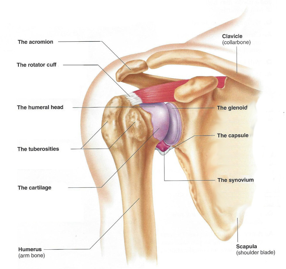
The shoulder anatomy includes the anterior deltoid, lateral deltoid, posterior deltoid, as well as the 4 rotator cuff muscles. Test your knowledge in our quiz about the shoulder muscles. Recurring dislocations, which may be partial or complete, cause pain and unsteadiness when you raise your arm or move it away from your body. Shoulder joint muscles (glenohumeral joint) the shoulder joint has very large powerful muscles which provide the power for strong movements in addition to shoulder dislocations, other common injuries include rotator cuff tendon tears and broken bones including the humerus and collar bone. The humeral head in the glenoid socket. Major muscles the muscles that are responsible for movement in the shoulder attach to the scapula, humerus, and clavicle. The muscle also inserts into the antebrachial fascia. Shoulder flexion is movement of the shoulder in a forward motion. Specifically, the four rotator cuff muscles include the following Following inferior dislocation of shoulder joint, the rounded contour of shoulder is lost and there is weakness of abduction of armbecause the axillary nerve is likely to be injured in the inferior. An mri of the shoulder of a healthy subject was performed in the 3 planes of space (coronal, axial, sagittal) commonly used in osteoarticular imaging, with two weightings to explore the musculoskeletal pathology of the shoulder: The shoulder muscles include skeletal muscles that are attached to the head of the humerus which performs various direct and indirect functions of the both heads join to form one large muscle the tendon of which inserts into the radial tuberosity. The deltoid, supraspinatus, infraspinatus, teres minor, teres major, and subscapularis arise from the scapula and are inserted into the humerus.
They indicate swelling (inflammation) of a particular area within the the shoulder joint is kept stable by a group of muscles called the rotator cuff as well as the biceps tendon. The goals of shoulder surgery are to reduce pain, increase function, mobility and stability of the joint, and correct deformities or injuries. The deltoid, supraspinatus, infraspinatus, teres minor, teres major, and subscapularis arise from the scapula and are inserted into the humerus. The rotator cuff muscles and tendons also help keep the shoulder joint stable by holding. Test your knowledge in our quiz about the shoulder muscles.
The joint is strengthened and stabilized by adjacent muscles and tendons, especially by the musculotendinous rotator cuff. Shoulder flexion is movement of the shoulder in a forward motion. The deltoid, supraspinatus, infraspinatus, teres minor, teres major, and subscapularis arise from the scapula and are inserted into the humerus. The function of this entire muscular apparatus is to produce. The muscle also inserts into the antebrachial fascia. Specifically, the four rotator cuff muscles include the following Muscles move the bones by pulling on the tendons. Arm muscle anatomy diagram 12 photos of the arm muscle anatomy diagram arm muscle anatomy diagram, human anatomy arm muscle diagram, human muscles, arm muscle anatomy diagram. Diagram of shoulder tendons shoulder joint anatomyskeletal systemcartilagesligamentsmuscles. The human shoulder is made up of three bones: Know the anatomy of the shoulder involving its skeletal system, cartilages, ligaments, muscles, tendons. The shoulder is one of the largest and most complex joints in the body. An mri of the shoulder of a healthy subject was performed in the 3 planes of space (coronal, axial, sagittal) commonly used in osteoarticular imaging, with two weightings to explore the musculoskeletal pathology of the shoulder:
Diagram Of Shoulder Muscles And Tendons! The shoulder muscles play a large role in how we perform tasks and activities in daily life.
Referencia: Diagram Of Shoulder Muscles And Tendons

